13 Skeletal System
Topic: Skeletal System
Text Reference: Chapter 13. Skeletal System
Objectives: Students should be able to…
Identify meanings of key word components of the musculoskeletal system
Prefixes
a- (absence of, without)
ab- (away from)
ad- (towards)
brady- (slow)
dys- (painful, difficult, abnormal, labored)
hyper- (above, excessive)
inter- (between)
intra- (within, in)
poly- (many, much)
sub- (below, under)
supra- (above)
sym- (together, joined)
syn- (together, joined)
Combining Forms
ankyl/o (stiff, bent)
aponeur/o (aponeurosis)
arthr/o (joint)
burs/o (bursa)
carp/o (carpals, wrist)
chondr/o (cartilage)
clavic/o (clavicle, collarbone)
clavicul/o (clavicle, collarbone)
cost/o (ribs)
crani/o (cranium)
disk/o (intervertebral disk)
femor/o (femur, upper leg bone)
fibul/o (fibula, lower leg bone)
humer/o (humerus, upper arm bone)
ili/o (ilium)
ischi/o (ischium)
kinesi/o (movement, motion)
kyph/o (increased convexity of the spine)
lord/o (bent forward, increased concavity of the spine)
lumb/o (loin, lumbar region of the spine)
mandibul/o (mandible, lower jaw bone)
maxill/o (maxilla, upper jaw bone)
menisc/o (meniscus, crescent)
myel/o (marrow [bone], spinal cord)
oste/o (bone)
patell/o (patella, kneecap)
pelv/i (pelvis, pelvic bone)
pelv/o (pelvis, pelvic bone)
petr/o (stone)
phalang/o (phalanges, bones of finger and toes)
pub/o (pubis)
rachi/o (vertebral spine, vertebral column)
radi/o (nerve root)
scapul/o (scapula, shoulder blade)
scoli/o (crooked, curved)
spondyl/o (vertebra, spine, vertebral column)
stern/o (sternum, breast bone)
tars/o (tarsals, ankle bones)
ten/o (tendon)
tendin/o (tendon)
tend/o (tendon)
tibi/o (tibia, lower leg bone)
uln/o (ulna, lower arm bone)
vertebr/o (vertebra, spine, vertebral column)
Suffixes
-al (pertaining to)
-algia (pain)
-ar (pertaining to)
-asthenia (weakness)
-centesis (surgical puncture to aspirate fluid)
-clasia (break)
-clasis (break)
-clast (break)
-desis (surgical fixation, fusion)
-ectomy (excision, surgical removal, cutting out)
-gram (the record, radiographic image)
-graphy (process of recording, radiographic imaging)
-ic (pertianing to)
-itis (inflammation)
-lysis (loosening, separating, dissolution)
-malacia (softening)
-oid (resembling)
-oma (tumor)
-osis (abnormal condition)
-penia (abnormal reduction)
-physis (growth)
-plasty (surgical repair)
-rrhaphy (suturing, repairing)
-sarcoma (malignant tumour)
-schisis (split, fissure)
-scopy (process of viewing, visual examination)
-tomy (incision, cut into)
-trophy (nourishment, development)
Apply the rules of medical language to pronounce, break into word parts, and define the following terms.
Label each word part by using the following abbreviations:
P = Prefix
WR = Word Root
CV = Combining Vowel
S = Suffix
CF = Combining Form
Example: osteoarthropathy (ä-stē-ō-är-THROP-ă-thē) – disease of bone and joint
WR CV WR CV S
oste / o / arthr / o /pathy
CF CF
Practice pronouncing and defining these musculoskeletal system movement terms.
abduction (ab-DŬK-shŏn)
adduction (ă-DŬK-shŏn)
eversion (ē-VĔR-zhŭn)
extension (ek-STEN-shŏn)
flexion (FLEK-shŏn)
inversion (in-VĔR-zhŭn)
pronation (prō-NĀ-shŭn)
rotation (rō-TĀ-shŏn)
supination (sū-pĭn-Ā-shŭn)
Sort the terms from the word lists above into the following categories.
- Disease and Disorder (terms describing any deviation from normal structure and function)
- Diagnostic (terms related to process of identifying a disease, condition, or injury from its signs and symptoms)
- Therapeutic (terms related to treatment or curing of diseases)
- Anatomic (terms related to body structure)
Use terms related to the musculoskeletal system.
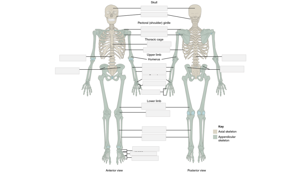
Label the following skeletal system anatomy.
carpals | clavicle | cranial portion | facial portion | femur | fibula | humerus | metacarpals | metatarsals | patella | pelvic girdle | phalanges | phalanges | pelvic girdle | radius | ribs | scapula | sternum | tarsals | tibia | ulna | vertebral column | vertebral column
Place the following medical terms in context to complete the scenario below.
arthritis | arthroscopy | chondromalacia | femoral | Orthopedic | tendinitis | total hip arthroplasty | x-rays
MUSCULOSKELETAL SYSTEM – OPERATIVE REPORT
PATIENT NAME: Mrs. Karen SMITH
AGE: 72 Sex: Female
DATE OF SURGERY: February 24
PREOPERATIVE DIAGNOSIS: Degenerative arthritis of both hips; more severe on the right side.
POSTOPERATIVE DIAGNOSIS: Severe degenerative arthritis and severe ________ of the right hip.
NAME OF PROCEDURE: Total hip arthroplasty.
HISTORY: Mrs. Karen Smith is a 72-year-old widow who has been living alone and independently since her husband’s premature death 15 years ago. Mrs. Smith has worked for 30 years at the production line in a factory and is now retired.
Mrs. Smith has been experiencing discomfort in her hips, especially the right one, over the parts twenty years or so. However, what started as a mild discomfort over time turned into severe pain. Now the pain is so bad that she is afraid that it might soon rob her of her independence. She first sought help for her hip pain many years ago. After physical examination, her family physician ordered ________ for both hip joints. Based on the results, it was concluded that the pain was due to severe ________ mainly due to wear and tear. She was advised to lose weight and to take over-the-counter painkillers as needed. She was also referred to a physiotherapist. However, despite the fact that she has lost 10% of her original body weight of 170 pounds and has been adhering to the exercise regimen recommended by her physiotherapist, the pain has grown worse over the years and now is almost unbearable. She was last visited by an orthopedic surgeon and subsequently was admitted to the General Hospital Outpatient ________ Clinic for ________ of both hips.
OPERATIVE REPORT: The patient was brought to the operating room by anesthesia personnel. She was placed on the operating table. A Foley catheter was inserted. The patient was then placed into the lateral decubitus position with her right side up. The right lower extremity was prepped and draped in standard fashion for a ________. Dissection was carried sharply down through the soft tissue to the greater trochanter. The greater trochanter was used as a landmark to orient the remainder of the dissection which was continued posteriorly and proximally to expose the iliofemoral joint.
The acetabulum was reamed. A 50 mm acetabular shell was used. Femur was debrided using a ________ canal curette. The length of the femoral stem was then checked with the canal curette in place. Appropriate femoral stem and head were selected and implanted. Intraoperative radiographs were obtained to ensure proper component position.
The hip was then checked for range of motion. The patient reached 90 degrees of flexion and full extension with no instability. No abnormality was detected in the surrounding soft tissue. There was no indication of ________.
The area was then closed in a layered fashion. The subcutaneous tissues were closed using surgical Vicryl 5-0 sutures. An incisional VAC was placed over the wound as well. Sponge and needle counts were correct at the end of the operation. The patient tolerated the procedure well and was returned to the recovery room in good condition.
__________________________________
Michael Porter, MD, Orthopedic Surgery
Place the following medical terms in context to complete the scenario below.
akinesia | aligned | arthralgia | atrophy | carpal | Colles | fluoroscopy | orthopedic | splint | sterilized | supination | sutured | tenomyoplasty | tenorrhaphy
MUSCULOSKELETAL SYSTEM – OPERATIVE REPORT
PATIENT NAME: Liam PALMER
AGE: 22
SEX: Male
DOB: December 4
DATE OF ADMISSION: May 5
DATE OF PROCEDURE: May 5
ATTENDING PHYSICIAN: Michael Porter, MD, Orthopedic Surgery
PREOPERATIVE DIAGNOSIS: Fx of the distal end of radius.
POSTOPERATIVE DIAGNOSIS: Fx of the distal end of radius.
ANESTHESIA: General.
INDICATION: This 22-year-old male had been skating earlier today when he lost his balance and fell. Trying to break the fall with an outstretched arm, he landed on his right arm, breaking his wrist. Mr. Palmer was brought to the ________ clinic in Toronto General Hospital. The wrist has been kept in a neutral position since even a slight movement was painful. The injured area is edematous and any attempt for active or passive flexion, extension, ________, or pronation caused a sharp pain that shoots all the way to the right shoulder. Posterior-Anterior and lateral x-rays of the wrist and forearm confirmed ________ fracture of the distal end of radius with the broken piece displaced posteriorly. The ________ bones were intact. The patient required surgery to fix the broken bone. Although not certain at that point, there was a possibility that the patient also required ________.
PROCEDURE: The surgery was done under general anesthesia. The patient’s arm was placed in a proper position to allow for an easy and unobstructed access to the surgical area. The surgical area was ________. A longitudinal incision was made to access the fracture. The fractured bone was realigned, and a metal plate was used to secure the ________ bone and restore stability. Throughout the surgery ________ was used to ensure proper reduction of the bone. The surrounding muscles, tendons, and ligaments were examined to ensure their integrity. There was no need for tenomyoplasty or ________. Once the surgery was completed, the surgical incision was ________, the wrist was bandaged, and the arm was placed in a long cast to immobilize the wrist and elbow joints. The patient left the operation room in good and stable condition.
The patient was discharged from the hospital on the following day. He was scheduled for his first follow up visit in 3 weeks. At that time, the cast will be replaced with a removable wrist ________ and the patient will be referred to a physiotherapy clinic. Timely rehabilitation is extremely important in these types of fractures to reduce ________ and prevent from ________ and muscle ________.
_________________________________________
Michael Porter, MD, Orthopedic Surgery
Test your knowledge by answering the questions below.
Joints with some movement are called…
- Synarhrosis
- Amphiarthrosis
- Diarthrosis
The skeleton that consists of all the bones in the upper and lower limbs is called…
- Axial Skeleton
- Articulations
- Appendicular Skeleton
A condition that lasts a long time with periods of remission and exacerbation is called…
- Chronic
- Hematopoiesis
- Edema
Forward curvature of the lower lumbar spine is called…
- Lordosis
- Kyphosis
- Scoliosis
Comminuted fractures are…
- Bones that are broken and pierce through the skin
- Bones that are broken and crushed in pieces
- Bones that are broken but do not protrude the skin
