Integumentary
10.7 Putting It All Together
Review the following example of applying the nursing process to a patient with a pressure injury.
Patient Scenario

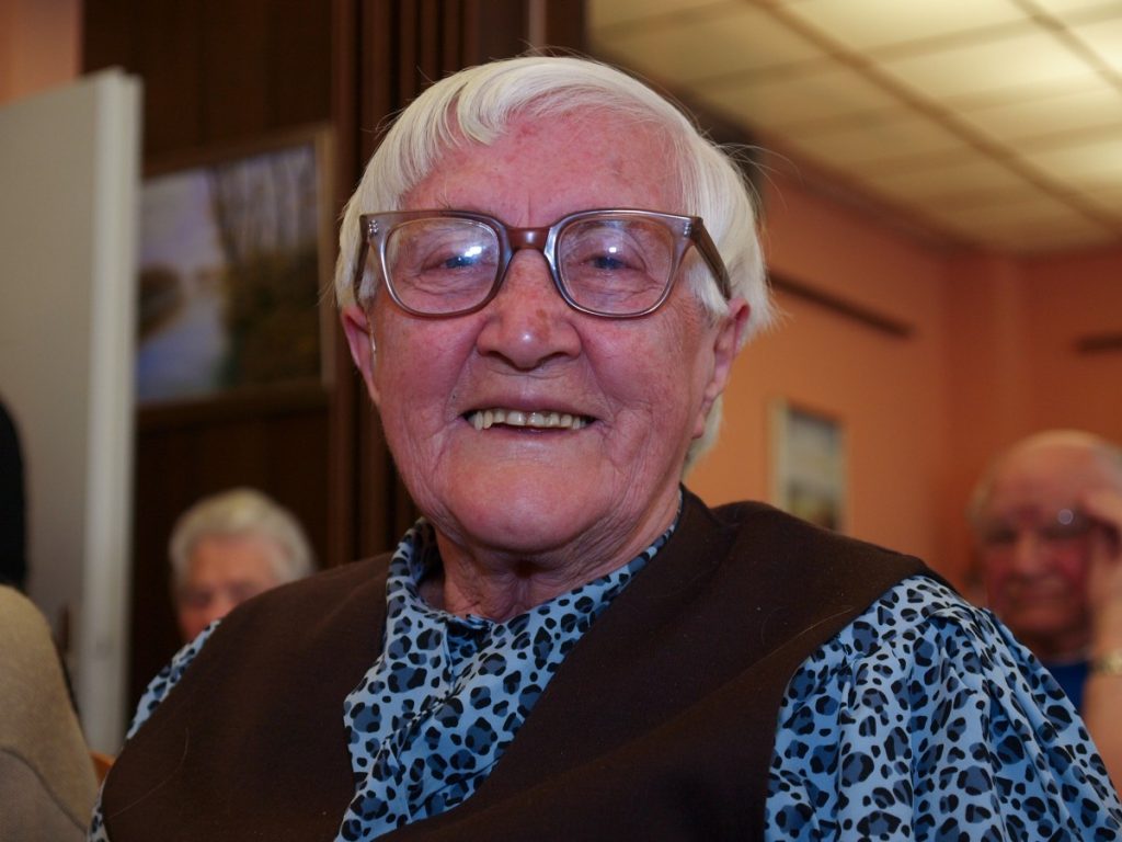
Betty Pruitt is a 92-year-old female admitted to a skilled nursing facility after a fall at her daughter’s home while transferring the patient from her bed to a wheelchair. See Figure 10.24 for an image of Betty Pruitt.[1]Although no injury was sustained, it became clear to the family that they could no longer provide adequate care at home.
Ms. Pruitt’s past medical history includes congestive heart failure, hypertension, hypercholesterolemia, and moderate stage Alzheimer’s disease. Her cognitive ability has significantly declined over the last six months. Patient’s speech continues to be mostly clear and at times coherent but she tends to be quiet and does not express her needs adequately, even with prompting. She no longer has the ability to ambulate but can stand for short periods of time, requiring two people to transfer. She rarely changes body position without encouragement and assistance, spending most of her days in a recliner or bed. Betty is 69 inches tall and currently weighs 122 pounds, having lost 22 pounds over the last 3 months. BMI is 18. Family reports her appetite is poor, and she eats only in small amounts at meal times with feeding assistance. She does take liquids well and shows no swallowing difficulties at this time. Betty is incontinent of urine and stool most of the time but will use the toilet if offered and given transfer help. Unknown to the family, a skin assessment revealed a Stage III pressure injury on coccyx area. Wound measures 4 cm long, 4 cm wide, 3 cm deep, with adipose tissue visible. No undermining, tunneling, bone, muscle, or tendons visible. Scant amount of yellowish purulent drainage noted. Slight foul odor, with redness, and increased heat around the wound present.
A Braden Scale Risk Assessment was completed and revealed a total score of 12 (High Risk) with the following category scores: Sensory Perception-3, Moisture-2, Activity-2, Mobility-2, Nutrition-2, Friction & Shear-1.
Applying the Nursing Process
Based on this information, the following nursing care plan was implemented for Ms. Pruitt.
Nursing Diagnosis: Impaired Tissue Integrity related to imbalanced nutritional state and associated with impaired mobility as evidenced by damaged tissue, redness, area hot to touch.
Overall Goal: The patient will experience wound healing demonstrated by decreased wound size and increased granulation tissue.
SMART Expected Outcome: Ms. Pruitt will have a 50% reduction in wound dimensions (from 4 cm in diameter to 2 cm) within two weeks.
Planned Nursing Interventions with Rationale: See Table 10.7 for a list of planned nursing interventions with rationale.
Table 10.7 Selected Interventions and Rationale for Ms. Pruitt
| Interventions | Rationale |
|---|---|
| 1. Assess and document wound characteristics every shift, including size (length x width x depth), stage (I-IV), location, exudate, presence of granulation tissue, and epithelization. | Consistent and accurate documentation of wounds is important in determining the progression of wound healing and effectiveness of treatments. |
| 2. Monitor for signs of infection (color, temperature, edema, moisture, pain, and appearance of surrounding skin). | Frequent monitoring for possible wound infection provides the ability to intervene quickly if changes in the wound are noted. Additionally, pain medications should be offered prior to dressing changes if pain is present. |
| 3. Cleanse wound per facility protocol or as ordered. | Removal of exudate, dirt, and slough promotes wound healing. |
| 4. Cleanse the periwound area (skin around the wound) with mild soap and water. | Decreasing the number of microorganisms around the wound may decrease the chance of wound infection. |
| 5. Apply and change wound dressings, per facility protocol or wound orders. | Dressings that maintain moisture in the wound keep periwound skin dry, absorb drainage, and pad the wound to protect from further injury assist in healing. |
| 6. Turn/reposition the patient every 2 hours and position with pillows as needed. | Frequent repositioning relieves pressure point areas from damage. Avoid positioning the patient directly on an injured area if possible. |
| 7. Consider the use of a specialty mattress, bed, or chair pad. | Specialty mattresses, beds, or pads offer added padding and support, while decreasing pressure areas. |
| 8. Use moisture barrier ointments (protective skin barriers). | Moisture barrier ointments can significantly decrease skin breakdown and pressure injury formation. |
| 9. Check incontinence pads frequently (every 2-3 hours) and change as needed to keep dry. | Frequent changing of soiled pads will prevent exposure to chemicals in urine and stool that erode the skin. |
| 10. Monitor nutritional status and obtain order for dietary consult if needed. | Optimizing nutritional intake, including calories, protein, and vitamins, is essential to promote wound healing. |
| 11. Offer nutritional supplements and water. | Nutritional supplements, such as protein shakes, can provide additional calories and protein without a large volume of intake needed. Water intake is essential for proper tissue hydration. |
| 12. Keep bed linens clean, dry, and wrinkle free. | Soiled, wet, or wrinkled sheets may contribute to skin breakdown. |
| 13. Use a minimum of two-person assistance and a draw sheet to pull the patient up in bed. | Carefully transferring patients avoids adverse effects of external mechanical forces (pressure, friction, and shear) from causing skin or tissue damage. |
Interventions Implemented:
After the admission assessment was completed, Ms. Pruitt became settled in her new room. The wound was assessed, documented, and cleaned. A specimen for wound culture was obtained and a wound dressing applied per protocol. The health care provider was notified of the wound. Requests were made for a wound culture, referrals to a wound care nurse specialist and a dietician, and a pressure-relieving mattress for the bed. A two-hour turning schedule was implemented, and the CNA was reminded to use two-person assistance with a lift sheet when repositioning the patient. A barrier cream was applied to protect the peri-area whenever a new incontinence pad was placed. The following documentation note was entered in the patient chart.
Documentation:
On admission, a Stage III pressure injury was discovered on the patient’s coccyx area. The wound measured 4 cm long, 4 cm wide, 3 cm deep, with adipose tissue visible. No undermining, tunneling, bone, muscle, or tendons visible. A small amount of yellow purulent drainage noted. Slight foul odor, with redness, and increased heat around the wound present. Wound was cleaned with normal saline and packed with moist gauze and covered with hydrogel dressing. Patient tolerated the procedure well and gave no evidence of pain. A pressure-relieving mattress was placed on the patient’s bed and a two-hour turning schedule was implemented. Patient voided x 1 and the pad was changed. Barrier cream was applied to the perineal area. Patient encouraged to rest until lunchtime and is resting.
Evaluation: After two weeks, the measurements of the wound were compared to those on admission and the wound decreased in size to less than 2 cm. The expected outcome was “met.” A new expected outcome was established, “Mrs. Pruitt’s wound will resolve within the next 2 weeks.” The same planned interventions were continued to be implemented.
- “1068481.jpg” by unknown is licensed under CC0 ↵

