6.2 Basic Neurological Concepts
Open Resources for Nursing (Open RN)
When completing a neurological assessment, it is important to understand the functions performed by different parts of the nervous system while analyzing findings. For example, damage to specific areas of the brain, such as that caused by a head injury or cerebrovascular accidents (i.e., strokes), can cause specific deficits in speech, facial movements, or use of the extremities. Damage to the spinal cord, such as that caused by a motor vehicle accident or diving accident, will cause specific motor and sensory deficits according to the level where the spinal cord was damaged.
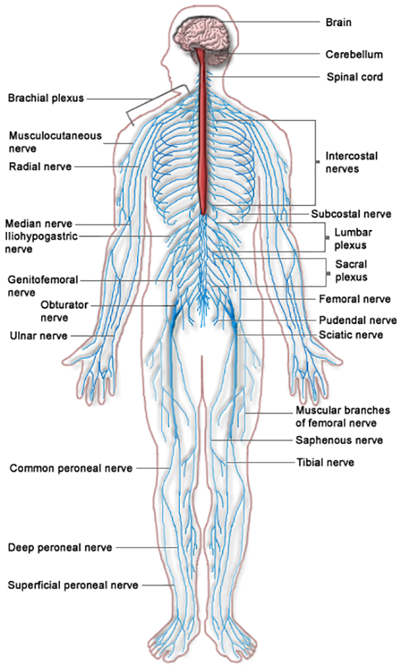
The nervous system is divided into two parts, the central nervous system and the peripheral nervous system. See Figure 6.1[1]for an image of the entire nervous system. The central nervous system (CNS) includes the brain and the spinal cord. The brain can be described as the interpretation center, and the spinal cord can be described as the transmission pathway. The peripheral nervous system (PNS) consists of the neurological system outside of the brain and spinal cord, including the cranial nerves that branch out from the brain and the spinal nerves that branch out from the spinal cord. The peripheral nervous system can be described as the communication network between the brain and the body parts. Both parts of the nervous system must work correctly for healthy body functioning.
Central Nervous System
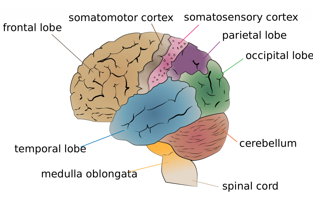
There major regions of the brain are the cerebrum and cerebral cortex, the diencephalon, the brain stem, and the cerebellum. See Figure 6.2[2] for an illustration of the cerebellum and the lobes of the cerebrum.
Cerebrum and Cerebral Cortex
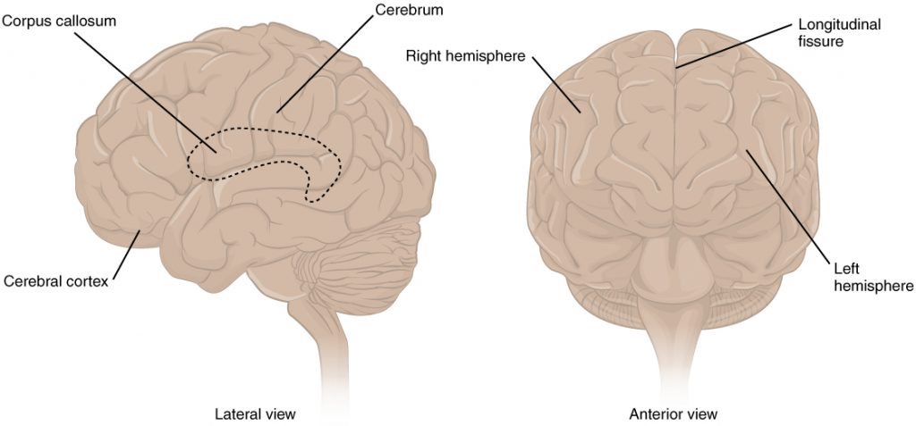
The largest portion of our brain is the cerebrum. The cerebrum is covered by a wrinkled outer layer of gray matter called the cerebral cortex. See Figure 6.3[3] for an image of the cerebral cortex. The cerebral cortex is responsible for the higher functions of the nervous system such as memory, emotion, and consciousness. The corpus callosum is the major pathway of communication between the right and left hemispheres of the cerebral cortex. The cerebral cortex is further divided into four lobes named the frontal, parietal, occipital, and temporal lobes.[4] Each lobe has specific functions.
Frontal Lobe
The frontal lobe is associated with movement because it contains neurons that instruct cells in the spinal cord to move skeletal muscles. The anterior portion of the frontal lobe is called the prefrontal lobe, and it provides cognitive functions such as planning and problem-solving that are the basis of our personality, short-term memory, and consciousness. Broca’s area is also located in the frontal lobe and is responsible for the production of language and controlling movements responsible for speech.[5]
Parietal Lobe
The parietal lobe processes general sensations from the body. All of the tactile senses are processed in this area, including touch, pressure, tickle, pain, itch, and vibration, as well as general senses of the body, such as proprioception (the sense of body position) and kinesthesia (the sense of movement).[6]
Temporal Lobe
The temporal lobe processes auditory information and is involved with language comprehension and production. Wernicke’s area and Broca’s area are located in the temporal lobe. Wernicke’s area is involved in the comprehension of written and spoken language, and Broca’s area is involved in the production of language. Because regions of the temporal lobe are part of the limbic system, memory is also an important function associated with the temporal lobe.[7] The limbic system is involved with our behavioral and emotional responses needed for survival, such as feeding, reproduction, and the fight – or – flight responses.
Occipital Lobe
The occipital lobe primarily processes visual information.[8]
Diencephalon
Information from the rest of the central and peripheral nervous system is sent to the cerebrum through the diencephalon, with the exception of the olfactory nerve that connects directly to the cerebrum.[9] See Figure 6.4[10] for an illustration of the diencephalon deep within the cerebrum. The diencephalon contains the hypothalamus and the thalamus.
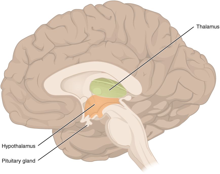
The hypothalamus helps regulate homeostasis such as body temperature, thirst, hunger, and sleep. The hypothalamus is also the executive region in charge of the autonomic nervous system and the endocrine system through its regulation of the anterior pituitary gland. Other parts of the hypothalamus are involved in memory and emotion as part of the limbic system.[11]
The thalamus relays sensory information and motor information in collaboration with the cerebellum. The thalamus does not just pass the information on, but it also processes and prioritizes that information. For example, the portion of the thalamus that receives visual information will influence what visual stimuli are considered important enough to receive further attention from the brain.[12]
Brain Stem
The brain stem is composed of the pons and the medulla. The pons and the medulla regulate several crucial autonomic functions in the body, including involuntary functions in the cardiovascular and respiratory systems, vasodilation, and reflexes like vomiting, coughing, sneezing, and swallowing. Cranial nerves also connect to the brain through the brain stem and provide sensory input and motor output.[13]
Cerebellum
The cerebellum is located in the posterior part of the brain behind the brain stem and is responsible for fine motor movements and coordination. For example, when the motor neurons in the frontal lobe of the cerebral cortex send a command down the spinal cord to initiate walking, a copy of that instruction is also sent to the cerebellum. Sensory feedback from the muscles and joints, proprioceptive information about the movements of walking, and sensations of balance are sent back to the cerebellum. If the person becomes unbalanced while walking because the ground is uneven, the cerebellum sends out a corrective command to compensate for the difference between the original cerebral cortex command and the sensory feedback.[14]
Spinal Cord
The spinal cord is a continuation of the brain stem that transmits sensory and motor impulses. The length of the spinal cord is divided into regions that correspond to the level at which spinal nerves pass through the vertebrae. Immediately adjacent to the brain stem is the cervical region, followed by the thoracic, the lumbar, and finally the sacral region.[15] The spinal nerves in each of these regions innervate specific parts of the body. See more information under the “Spinal Nerves” section.
Review the anatomy of the brain using following supplementary video.
Video Review for Anatomy of the Brain[16]
Peripheral Nervous System
The peripheral nervous system (PNS) consists of cranial nerves and spinal nerves that exist outside of the brain, spinal cord, and autonomic nervous system. The main function of the PNS is to connect the limbs and organs to the central nervous system (CNS). Sensory information from the body enters the CNS through cranial and spinal nerves. Cranial nerves are connected directly to the brain, whereas spinal nerves are connected to the brain via the spinal cord.
Peripheral nerves are classified as sensory nerves, motor nerves, or a combination of both. Sensory nerves carry impulses from the body to the brain for processing. Motor nerves transmit motor signals from the brain to the muscles to cause movement.
Cranial Nerves
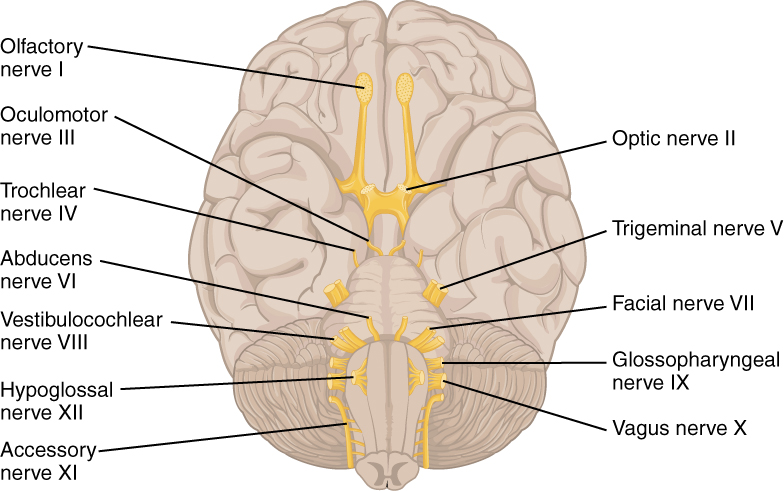
Cranial nerves are directly connected from the periphery to the brain. They are primarily responsible for the sensory and motor functions of the head and neck. There are twelve cranial nerves that are designated by Roman numerals I through XII. See Figure 6.5[17] for an image of cranial nerves. Three cranial nerves are strictly sensory nerves; five are strictly motor nerves; and the remaining four are mixed nerves.[18] A traditional mnemonic for memorizing the names of the cranial nerves is “On Old Olympus Towering Tops A Finn And German Viewed Some Hops,” in which the initial letter of each word corresponds to the initial letter in the name of each nerve.
- The olfactory nerve is responsible for the sense of smell.
- The optic nerve is responsible for the sense of vision.
- The oculomotor nerve regulates eye movements by controlling four of the extraocular muscles, lifting the upper eyelid when the eyes point up and for constricting the pupils.
- The trochlear nerve and the abducens nerve are both responsible for eye movement, but do so by controlling different extraocular muscles.
- The trigeminal nerve regulates skin sensations of the face and controls the muscles used for chewing.
- The facial nerve is responsible for the muscles involved in facial expressions, as well as part of the sense of taste and the production of saliva.
- The accessory nerve controls movements of the neck.
- The vestibulocochlear nerve manages hearing and balance.
- The glossopharyngeal nerve regulates the controlling muscles in the oral cavity and upper throat, as well as part of the sense of taste and the production of saliva.
- The vagus nerve is responsible for contributing to homeostatic control of the organs of the thoracic and upper abdominal cavities.
- The spinal accessory nerve controls the muscles of the neck, along with cervical spinal nerves.
- The hypoglossal nerve manages the muscles of the lower throat and tongue.[19] Methods for assessing each of these nerves are described in the “Assessing Cranial Nerves” section.
Video Review of Cranial Nerves[20]
Spinal Nerves
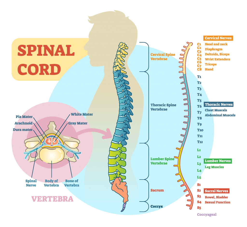
There are 31 spinal nerves that are named based on the level of the spinal cord where they emerge. See Figure 6.6[21] for an illustration of spinal nerves. There are eight pairs of cervical nerves designated C1 to C8, twelve thoracic nerves designated T1 to T12, five pairs of lumbar nerves designated L1 to L5, five pairs of sacral nerves designated S1 to S5, and one pair of coccygeal nerves. All spinal nerves are combined sensory and motor nerves. Spinal nerves extend outward from the vertebral column to innervate the periphery while also transmitting sensory information back to the CNS.[22]
Functions of Spinal Nerves
Each spinal nerve innervates a specific region of the body:
- C1 provides motor innervation to muscles at the base of the skull.[23]
- C2 and C3 provide both sensory and motor control to the back of the head and behind the ears.[24]
- The phrenic nerve arises from nerve roots C3, C4, and C5. This is a vital nerve because it innervates the diaphragm to enable breathing. If a patient’s spinal cord is transected above C3 from an injury, then spontaneous breathing is not possible.[25]
- C5 through C8 and T1 combine to form the brachial plexus, a tangled array of nerves that serve the upper limbs and upper back.[26]
- The lumbar plexus arises from L1-L5 and innervates the pelvic region and the anterior leg.[27]
- The sacral plexus comes from the lower lumbar nerves L4 and L5 and the sacral nerves S1 to S4. The most significant systemic nerve to come from this plexus is the sciatic nerve. The sciatic nerve is associated with the painful medical condition sciatica, which is back and leg pain as a result of compression or irritation of the sciatic nerve.[28]
Functions of the Nervous System
The nervous system receives information about the environment around us (sensation) and generates responses to that information (motor responses). The process of integration combines sensory perceptions and higher cognitive functions such as memories, learning, and emotion while producing a response.
Sensation
Sensation is defined as receiving information about the environment. The major senses are taste, smell, touch, sight, and hearing. Additional sensory stimuli are also provided from inside the body, such as the stretch of an organ wall or the concentration of certain ions in the blood.[29]
Response
The nervous system produces a response based on the stimuli perceived by sensory nerves. For example, withdrawing a hand from a hot stove is an example of a response to a painfully hot stimulus. Responses can be classified by those that are voluntary (such as contraction of a skeletal muscle) and those that are involuntary (such as contraction of smooth muscle in the intestine). Voluntary responses are governed by the somatic nervous system, and involuntary responses are governed by the autonomic nervous system.[30]
Integration
Integration occurs when stimuli received by sensory nerves are communicated to the nervous system and the information is processed, leading to the generation of a conscious response. Consider this example of sensory integration. A batter in a baseball game does not automatically swing when they see the baseball thrown to them by the pitcher. First, the trajectory of the ball and its speed will need to be considered before creating the motor response of a swing. Then, integration will occur as the batter generates a conscious decision of whether to swing or not. Perhaps the count is three balls and one strike, and the batter decides to let this pitch go by in the hope of getting a walk to first base. Perhaps the batter is afraid to strike out and doesn’t swing, or maybe the batter learned the pitcher’s nonverbal cues the previous time at bat and is confident to take a swing at an anticipated fast ball. All of these considerations are included as part of the batter’s integration response and the higher level functioning that occurs in the cerebral cortex.[31]
- “Nervous system diagram.png ” by unknown is licensed under CC BY-NC-SA 3.0. Access for free at https://med.libretexts.org/Bookshelves/Nursing/Book%3A_Clinical_Procedures_for_Safer_Patient_Care_(Doyle_and_McCutcheon)/02%3A_Patient_Assessment/2.07%3A_Focused_Assessments ↵
- “Cerebrum lobes.svg” by Jkwchui is licensed under CC BY-SA 3.0 ↵
- “1305 CerebrumN.jpg” by OpenStax is licensed under CC BY 4.0. Access for free at https://openstax.org/books/anatomy-and-physiology/pages/13-2-the-central-nervous-system ↵
- This work is a derivative of Anatomy & Physiology by OpenStax and is licensed under CC BY 4.0. Access for free at https://openstax.org/books/anatomy-and-physiology/pages/1-introduction ↵
- This work is a derivative of Anatomy & Physiology by OpenStax and is licensed under CC BY 4.0. Access for free at https://openstax.org/books/anatomy-and-physiology/pages/1-introduction ↵
- This work is a derivative of Anatomy & Physiology by OpenStax and is licensed under CC BY 4.0. Access for free at https://openstax.org/books/anatomy-and-physiology/pages/1-introduction ↵
- This work is a derivative of Anatomy & Physiology by OpenStax and is licensed under CC BY 4.0. Access for free at https://openstax.org/books/anatomy-and-physiology/pages/1-introduction ↵
- This work is a derivative of Anatomy & Physiology by OpenStax and is licensed under CC BY 4.0. Access for free at https://openstax.org/books/anatomy-and-physiology/pages/1-introduction ↵
- This work is a derivative of Anatomy & Physiology by OpenStax and is licensed under CC BY 4.0. Access for free at https://openstax.org/books/anatomy-and-physiology/pages/1-introduction ↵
- “1310 Diencephalon.jpg” by OpenStax is licensed under CC BY 4.0. Access for free at https://openstax.org/books/anatomy-and-physiology/pages/13-2-the-central-nervous-system ↵
- This work is a derivative of Anatomy & Physiology by OpenStax and is licensed under CC BY 4.0. Access for free at https://openstax.org/books/anatomy-and-physiology/pages/1-introduction ↵
- This work is a derivative of Anatomy & Physiology by OpenStax and is licensed under CC BY 4.0. Access for free at https://openstax.org/books/anatomy-and-physiology/pages/1-introduction ↵
- This work is a derivative of Anatomy & Physiology by OpenStax and is licensed under CC BY 4.0. Access for free at https://openstax.org/books/anatomy-and-physiology/pages/1-introduction ↵
- This work is a derivative of Anatomy & Physiology by OpenStax and is licensed under CC BY 4.0. Access for free at https://openstax.org/books/anatomy-and-physiology/pages/1-introduction ↵
- This work is a derivative of Anatomy & Physiology by OpenStax and is licensed under CC BY 4.0. Access for free at https://openstax.org/books/anatomy-and-physiology/pages/1-introduction ↵
- Forciea, B. (2015, May 12). Anatomy and physiology: Central nervous system: Brain anatomy v2.0. [Video]. YouTube. All rights reserved. Video used with permission. https://youtu.be/DBRdInd2-Vg ↵
- “1320 The Cranial Nerves.jpg” by OpenStax is licensed under CC BY 4.0. Access for free at https://openstax.org/books/anatomy-and-physiology/pages/13-4-the-peripheral-nervous-system ↵
- This work is a derivative of Anatomy & Physiology by OpenStax and is licensed under CC BY 4.0. Access for free at https://openstax.org/books/anatomy-and-physiology/pages/1-introduction ↵
- This work is a derivative of Anatomy & Physiology by OpenStax and is licensed under CC BY 4.0. Access for free at https://openstax.org/books/anatomy-and-physiology/pages/1-introduction ↵
- Forciea, B. (2015, May 12). Anatomy and physiology: Nervous system: Cranial nerves (v2.0). [Video]. YouTube. All rights reserved. Video used with permission. https://youtu.be/JBEZh6CHogo ↵
- "1008694237-vector.png" by VectorMine on Shutterstock. All rights reserved. Imaged used with purchased permission. ↵
- This work is a derivative of Anatomy & Physiology by OpenStax and is licensed under CC BY 4.0. Access for free at https://openstax.org/books/anatomy-and-physiology/pages/1-introduction ↵
- This work is a derivative of Anatomy and Physiology by Boundless.com and is licensed under CC BY-SA 4.0 ↵
- This work is a derivative of Anatomy and Physiology by Boundless.com and is licensed under CC BY-SA 4.0 ↵
- This work is a derivative of Anatomy and Physiology by Boundless.com and is licensed under CC BY-SA 4.0 ↵
- This work is a derivative of Anatomy and Physiology by Boundless.com and is licensed under CC BY-SA 4.0 ↵
- This work is a derivative of Anatomy & Physiology by OpenStax and is licensed under CC BY 4.0. Access for free at https://openstax.org/books/anatomy-and-physiology/pages/1-introduction ↵
- This work is a derivative of Anatomy & Physiology by OpenStax and is licensed under CC BY 4.0. Access for free at https://openstax.org/books/anatomy-and-physiology/pages/1-introduction ↵
- This work is a derivative of Anatomy & Physiology by OpenStax and is licensed under CC BY 4.0. Access for free at https://openstax.org/books/anatomy-and-physiology/pages/1-introduction ↵
- This work is a derivative of Anatomy & Physiology by OpenStax and is licensed under CC BY 4.0. Access for free at https://openstax.org/books/anatomy-and-physiology/pages/1-introduction ↵
- This work is a derivative of Anatomy & Physiology by OpenStax and is licensed under CC BY 4.0. Access for free at https://openstax.org/books/anatomy-and-physiology/pages/1-introduction ↵
The inability to raise the front part of the foot due to weakness or paralysis of the muscles that lift the foot.
The degree of range of motion a patient demonstrates in a joint when the examiner is providing the movement.
The thin, uppermost layer of skin.
The inner layer of skin with connective tissue, blood vessels, sweat glands, nerves, hair follicles, and other structures.
Sweat gland that produces hypotonic sweat for thermoregulation.
Sweat glands associated with hair follicles in densely hairy areas that release organic compounds subject to bacterial decomposition causing odor.
Skin pigment produced by melanocytes scattered throughout the epidermis.
A skin disturbance that typically occurs on areas of the skin that are rich in sebaceous glands, such as the face and back.
A tool used in the emergency department to assess the total body surface area burned to quickly estimate intravenous fluid requirements.
A type of swelling that occurs when lymph fluid builds up in the body's soft tissues due to damage to the lymph system.
A yellowing of the skin or sclera caused by underlying medical conditions.
Excessive, abnormal sweating.

